A Comprehensive Overview of the Functions of Human Bones (206)
Introduction
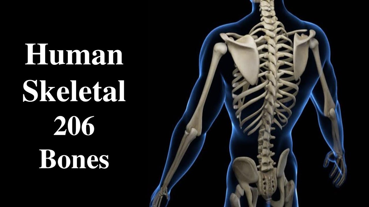
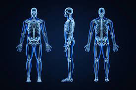
The human skeletal system serves as the framework of the body, providing structure, support, and protection to vital organs. Comprising 206 bones, each with its unique shape and function, the skeletal system is a marvel of engineering. From the sturdy long bones of the limbs to the delicate bones of the inner ear, every bone plays a crucial role in maintaining posture, facilitating movement, and safeguarding the body’s delicate tissues. In this comprehensive exploration, we delve into the intricate functions of each of the 206 human bones, illuminating the remarkable complexity and adaptability of the skeletal system.
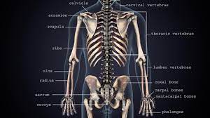
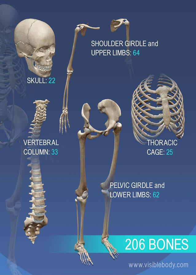
Skull: The skull, consisting of 22 bones, forms the protective enclosure for the brain and sensory organs. The cranium, comprising eight bones, encases the brain, providing it with vital protection against external trauma. Additionally, the facial bones, including the maxilla, mandible, and nasal bones, contribute to facial structure and house the sensory organs of sight, smell, and taste. The intricate arrangement of the skull bones also allows for the passage of nerves and blood vessels, facilitating essential physiological functions.
Vertebrae: The vertebral column, composed of 26 vertebrae, provides structural support and flexibility to the torso. Each vertebra is uniquely shaped to accommodate the spinal cord and nerves, allowing for the transmission of neural impulses between the brain and the rest of the body. Moreover, the intervertebral discs between the vertebrae act as shock absorbers, cushioning the spine and preventing injury during movement. Through its dynamic interplay of mobility and stability, the vertebral column enables an array of body movements, from bending and twisting to maintaining an upright posture.
Rib Cage: The rib cage, consisting of 24 ribs and the sternum, forms a protective enclosure around the thoracic cavity, safeguarding the heart, lungs, and major blood vessels. The ribs, with their curved structure, provide structural support to the chest while allowing for the expansion and contraction of the thoracic cavity during respiration. Additionally, the rib cage serves as an attachment site for muscles involved in breathing, posture, and upper limb movement, contributing to overall thoracic stability and mobility.
Upper Limbs: The upper limbs, comprising the clavicle, scapula, humerus, radius, ulna, carpals, metacarpals, and phalanges, facilitate a wide range of movements essential for daily activities and manual dexterity. The clavicle and scapula form the pectoral girdle, anchoring the upper limbs to the axial skeleton while allowing for shoulder mobility. The humerus, radius, and ulna articulate to form the elbow joint, enabling flexion, extension, and rotational movements of the forearm. The intricate arrangement of the carpals, metacarpals, and phalanges in the hand provides stability and precision grip, allowing for tasks ranging from fine motor skills to powerful grasping movements.
Pelvis: The pelvis, comprised of the ilium, ischium, pubis, and sacrum, forms a sturdy basin that supports the weight of the upper body and provides a stable foundation for the lower limbs. In addition to its role in weight-bearing and locomotion, the pelvis serves as a protective enclosure for the pelvic organs, including the reproductive organs and urinary bladder. The articulation of the pelvis with the femurs forms the hip joints, which facilitate a wide range of movements, from walking and running to sitting and squatting.
Lower Limbs: The lower limbs, consisting of the femur, patella, tibia, fibula, tarsals, metatarsals, and phalanges, are specialized for weight-bearing and locomotion. The femur, the longest and strongest bone in the body, forms the thigh and articulates with the pelvis to form the hip joint, transmitting the body’s weight during standing and walking. The patella, or kneecap, protects the knee joint and provides leverage for the quadriceps muscles during leg extension. The tibia and fibula articulate to form the ankle joint, supporting the body’s weight and providing stability during ambulation. The arrangement of the tarsals, metatarsals, and phalanges in the foot enables dynamic movements such as walking, running, jumping, and balancing, while also absorbing shock and maintaining posture.
Joints: Joints, or articulations, are points of contact between bones that allow for movement and flexibility. Classified based on their structure and function, joints range from immovable (synarthroses) to slightly movable (amphiarthroses) to freely movable (diarthroses). Synovial joints, such as the knee and shoulder joints, are characterized by their fluid-filled cavities and synovial membranes, which provide lubrication and reduce friction during movement. Cartilaginous joints, such as those between the vertebrae, are united by fibrocartilage or hyaline cartilage, allowing for limited movement while providing stability. Fibrous joints, such as those between the cranial bones, are held together by fibrous connective tissue and lack a joint cavity, providing maximum stability and protection to vital organs.
The human skeletal system comprises 206 bones, each with its unique structure and function. From providing structural support and protection to facilitating movement and locomotion, the bones of the body work in concert to maintain overall health and functionality. Through their remarkable adaptability and resilience, human bones embody the intricate balance between strength and flexibility, essential for the diverse range of activities that characterize human life. Understanding the functions of each bone not only deepens our appreciation for the complexity of the skeletal system but also underscores the interconnectedness of the human body as a whole.
Facial Bones: The facial bones, including the maxilla, mandible, nasal bones, zygomatic bones, and others, contribute to the structure and function of the face. These bones provide support for the soft tissues of the face, including the muscles responsible for facial expressions. Additionally, they house and protect the sensory organs of vision, smell, and taste. For example, the maxilla contains the sockets for the upper teeth and forms the floor of the nasal cavity, while the mandible supports the lower teeth and allows for chewing and speech production. The intricate arrangement of facial bones not only shapes individual facial features but also plays a crucial role in communication and social interaction.
Hyoid Bone: The hyoid bone, situated in the neck between the mandible and the larynx, serves as a point of attachment for muscles involved in swallowing and speech. Unlike other bones in the body, the hyoid bone does not articulate with any other bones but is suspended by ligaments and muscles. Its position and mobility are essential for the proper function of the tongue and larynx during swallowing and vocalization. Dysfunction of the hyoid bone can lead to swallowing difficulties, speech disorders, and other related issues, highlighting its importance in the complex processes of eating and communication.
Auditory Ossicles: The auditory ossicles, comprising the malleus, incus, and stapes, are the smallest bones in the human body and are located within the middle ear. These tiny bones play a crucial role in the transmission of sound vibrations from the tympanic membrane to the inner ear. As sound waves strike the tympanic membrane, it vibrates, causing the auditory ossicles to amplify and transmit these vibrations to the oval window of the inner ear. This process enables the conversion of sound waves into neural impulses, allowing for the perception of sound. The delicate structure and precise movements of the auditory ossicles are essential for normal hearing and auditory function.
Sesamoid Bones: Sesamoid bones are small, round bones embedded within tendons or joint capsules, where they act to reduce friction, increase leverage, and protect underlying tissues. The most well-known sesamoid bone in the human body is the patella, or kneecap, which lies within the tendon of the quadriceps muscle and functions to improve the mechanical advantage of knee extension. Additionally, sesamoid bones are found in the hands, feet, and other areas of the body where tendons pass over bony prominences, providing support and stability during movement. While sesamoid bones vary in size and location, they share the common function of enhancing musculoskeletal function and protecting vulnerable structures from excessive wear and tear.
Wormian Bones: Wormian bones, also known as sutural or intrasutural bones, are small, irregular bones that develop within the sutures of the skull. These additional bones arise from variations in ossification patterns and are often seen in individuals with certain genetic conditions or cranial deformities. While their exact function remains unclear, wormian bones are believed to contribute to the flexibility and resilience of the skull, particularly during childbirth and infancy when cranial bones are still forming. Although they are considered anatomical variations rather than distinct structures, wormian bones underscore the remarkable diversity and adaptability of the human skeletal system.
Supernumerary Bones: Supernumerary bones are additional skeletal structures that exceed the normal number of bones in a specific region of the body. These extra bones can occur as isolated anomalies or as part of genetic syndromes, developmental abnormalities, or acquired conditions. While supernumerary bones can be found in various parts of the skeleton, they most commonly affect the hands, feet, and spine. Depending on their location and size, supernumerary bones may or may not cause symptoms or functional impairments. Treatment options for supernumerary bones vary and may include observation, conservative management, or surgical intervention, depending on the individual’s clinical presentation and associated complications.
The human skeletal system is a complex and dynamic framework composed of 206 bones, each with its unique structure, function, and significance. From providing support and protection to facilitating movement, sensation, and communication, bones play a vital role in every aspect of human life. Understanding the functions of individual bones not only deepens our appreciation for the intricacies of the skeletal system but also highlights the remarkable adaptability and resilience of the human body. As ongoing research advances our knowledge of bone physiology and pathology, the significance of the skeletal system in health and disease continues to be an area of active investigation and clinical relevance.
Long Bones of the Extremities: Long bones, such as the femur, humerus, radius, and ulna, are characterized by their elongated shafts and relatively broad ends. These bones serve as levers for movement and are essential for supporting the body’s weight and facilitating locomotion. The cylindrical shape of long bones provides strength and rigidity while minimizing weight, making them well-suited for weight-bearing and load transmission. Additionally, the hollow medullary cavity within long bones contains bone marrow, where hematopoiesis, the production of blood cells, occurs. The dynamic interplay of cortical and trabecular bone tissue within long bones enables efficient energy absorption and distribution during physical activity, ensuring optimal performance and resilience.
Short Bones of the Hands and Feet: Short bones, such as the carpals in the hands and tarsals in the feet, are characterized by their cuboidal shape and compact structure. These bones provide stability and support to the wrist, hand, ankle, and foot joints while allowing for a wide range of movements. The articulation of short bones with adjacent bones and soft tissues forms intricate joint complexes that are essential for fine motor skills, balance, and proprioception. Additionally, the dense composition of short bones enhances their load-bearing capacity and resistance to compressive forces, making them integral components of the body’s weight-bearing architecture.
Irregular Bones of the Spine and Pelvis: Irregular bones, such as the vertebrae in the spine and the pelvic bones in the pelvis, have complex shapes and variable functions. These bones provide structural support and protection to vital organs while allowing for flexibility and mobility in the axial skeleton.
The vertebral column, comprised of irregularly shaped vertebrae, serves as the central axis of the body, supporting the head, neck, and trunk and providing attachment sites for muscles and ligaments. Similarly, the pelvic bones form a sturdy basin that supports the weight of the upper body and provides a stable foundation for the lower limbs. The unique morphology of irregular bones reflects their specialized roles in maintaining posture, facilitating movement, and protecting vulnerable structures within the body.
Sesamoid Bones of the Hands and Feet: Sesamoid bones, such as the patella in the knee and the pisiform in the hand, develop within tendons or joint capsules and serve to improve mechanical efficiency and reduce friction. These small, round bones act as pulleys or levers, enhancing the function of muscles and tendons during movement. For example, the patella increases the leverage of the quadriceps muscle during knee extension, while the pisiform provides support to the flexor tendons of the wrist and hand. The presence of sesamoid bones within weight-bearing joints also helps distribute forces more evenly, reducing the risk of injury and degeneration over time.
Axial Skeleton: The axial skeleton, composed of the skull, vertebral column, and rib cage, forms the central axis of the body and provides support and protection to vital organs. The skull encloses and protects the brain and sensory organs, while the vertebral column supports the head and trunk and allows for mobility and flexibility. The rib cage surrounds and protects the thoracic cavity, housing the heart, lungs, and major blood vessels. Together, these structures work in concert to maintain posture, facilitate movement, and safeguard the body’s delicate tissues, highlighting the integral role of the axial skeleton in overall health and functionality.
Appendicular Skeleton: The appendicular skeleton, comprised of the bones of the upper and lower limbs and their associated girdles, facilitates movement and locomotion and allows for interaction with the external environment. The pectoral girdle, consisting of the clavicle and scapula, anchors the upper limbs to the axial skeleton and provides attachment sites for muscles involved in shoulder movement and stability. The pelvic girdle, composed of the hip bones, supports the weight of the body and provides a stable foundation for the lower limbs. The bones of the upper and lower limbs articulate with one another to form complex joint systems that enable a wide range of movements, from fine motor skills to dynamic athletic activities.
Bone Marrow: Bone marrow, located within the medullary cavities of long bones and the spaces within spongy bone tissue, plays a crucial role in hematopoiesis, the production of blood cells. Red bone marrow, found predominantly in the axial skeleton and proximal ends of long bones, is responsible for generating red blood cells, white blood cells, and platelets.
These blood cells are essential for oxygen transport, immune function, and blood clotting, ensuring the body’s physiological integrity and homeostasis. Yellow bone marrow, found in the medullary cavities of long bones, consists primarily of adipocytes (fat cells) and serves as a reservoir of energy and nutrients. The dynamic regulation of bone marrow composition and function is essential for maintaining hematological health and systemic metabolic balance throughout life.
Conclusion
In conclusion, the human skeletal system is a marvel of biological engineering, comprising 206 bones that serve diverse functions essential for life and movement. From providing structural support and protection to facilitating movement and hematopoiesis, bones play integral roles in every aspect of human physiology and anatomy. Understanding the functions of individual bones and their interactions within the skeletal system illuminates the complexity and adaptability of the human body.
As ongoing research advances our knowledge of bone biology and pathology, the significance of the skeletal system in health and disease continues to be an area of active investigation and clinical relevance. Through interdisciplinary collaboration and technological innovation, we can further unravel the mysteries of the skeletal system and unlock new insights into human health and wellness.